Turning nuclear physics into medicine
A radiopharmaceutical is a tiny amount of a radioactive isotope, chemically guided to exactly where it is needed in the body. Get it right and you can see a tumour light up on a scan—or treat it from the inside, sparing healthy tissue. That is the promise this direction works to deliver.
Our Center for Radiopharmaceutical Research is a single, end-to-end infrastructure: a TR-19 cyclotron to create the isotopes, and a suite of shielded hot cells to turn them into sterile, injectable medicines. Around it we run fundamental, preclinical and clinical research that moves discoveries toward patients.
The journey of every dose follows three steps: make the isotope, see the disease, treat it.
1 · Make the isotope
From cyclotron beam to a vial that's safe to inject
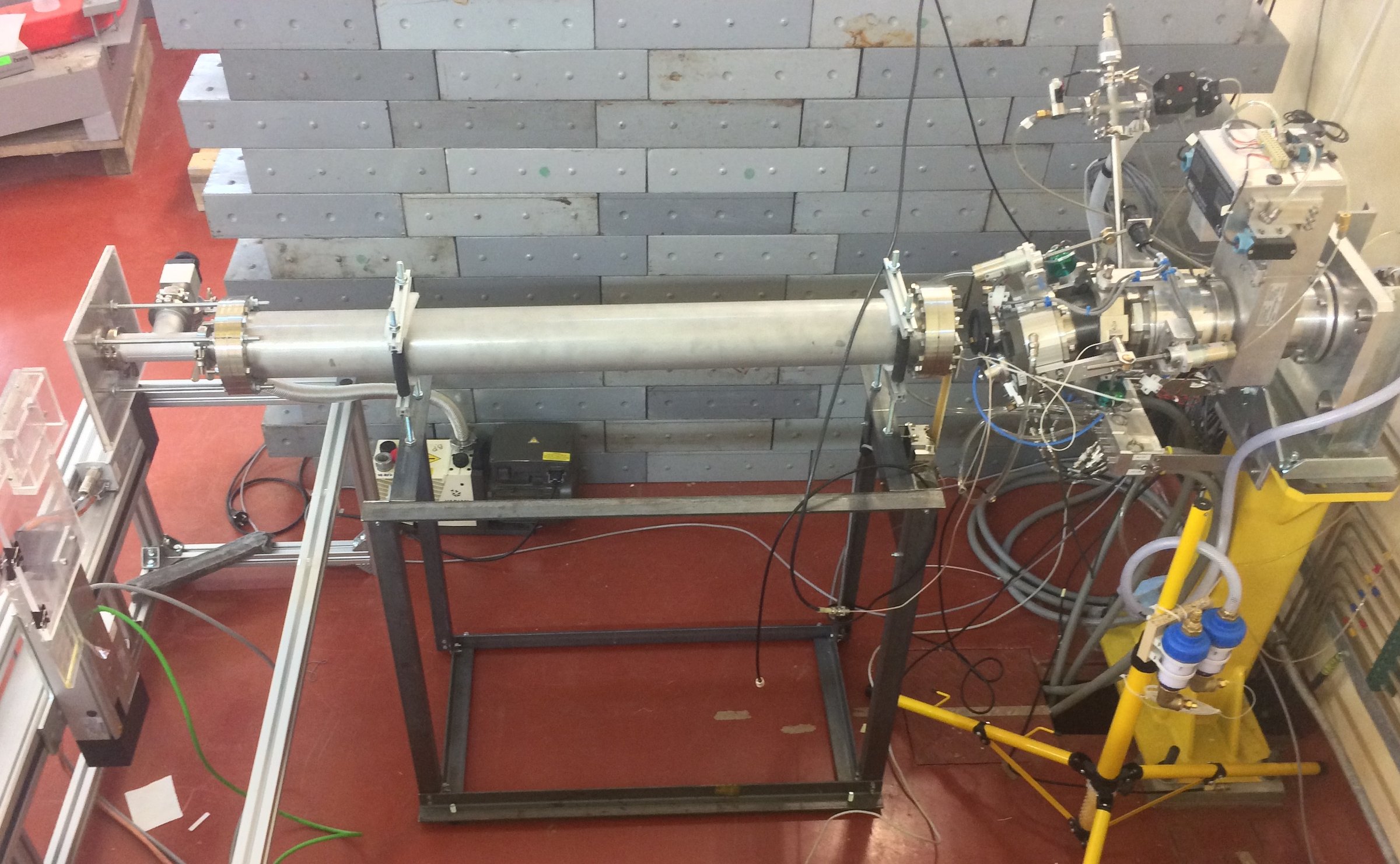
It starts with a target placed in the cyclotron's proton beam. The beam transmutes stable atoms into the short- and medium-lived radioisotopes that nuclear medicine depends on—diagnostic and therapeutic alike.
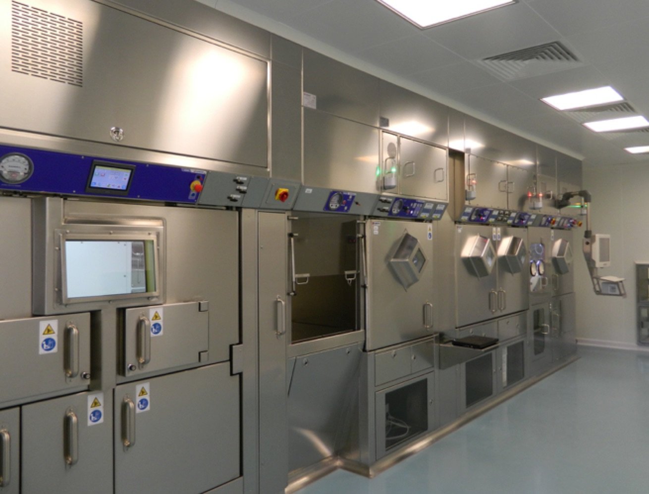
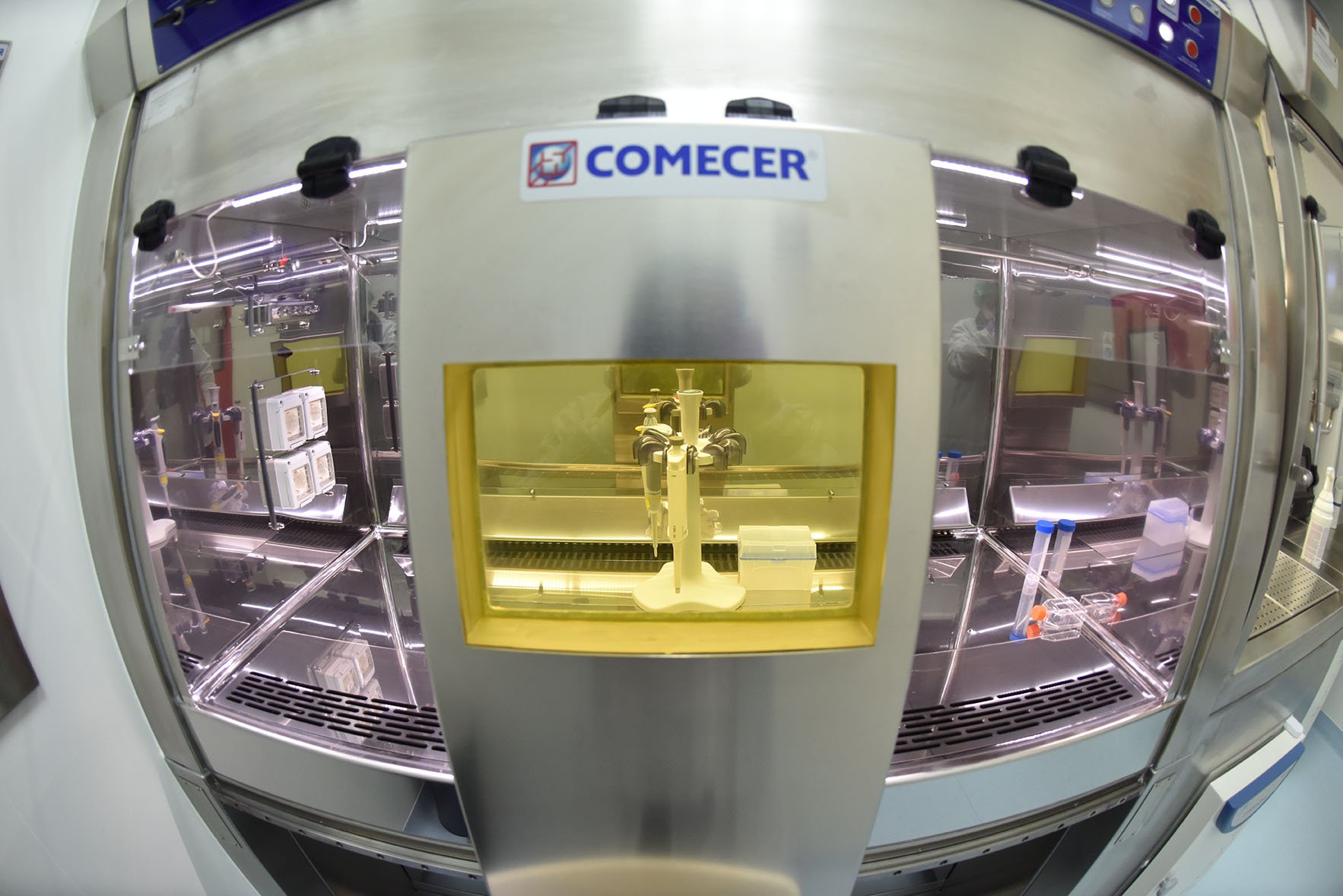
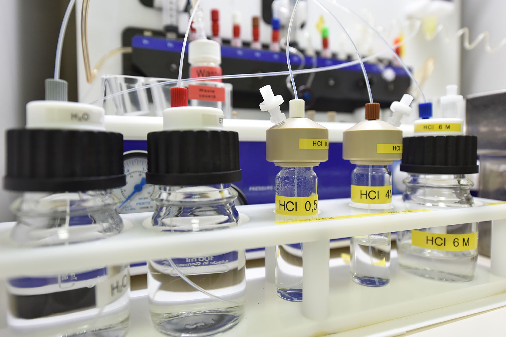
But a freshly irradiated target is not a medicine. Inside our shielded hot cells, the radioisotope is chemically separated, then linked to a targeting molecule that binds selectively to a specific receptor or biomarker in the diseased tissue, formulated under sterile conditions and rigorously quality-checked before it can ever reach a patient. We own this whole chain—targetry, irradiation, separation, synthesis, sterile formulation, testing and release—for both research batches and early clinical translation.
2 · See the disease
Molecular imaging that finds disease earlier
Once a radiopharmaceutical is in the body, it becomes a beacon. PET and SPECT cameras detect the radiation it emits and reconstruct where it has gone—revealing biological function: how tissues are metabolising, where a tumour is active, how an organ is working.
Because these images show biology rather than just structure, they can catch disease earlier and guide treatment more precisely than anatomy-only scans—especially when combined with CT or MR. This is the foundation of personalised nuclear medicine, and a core of our drive to build a centre of excellence in nuclear imaging.
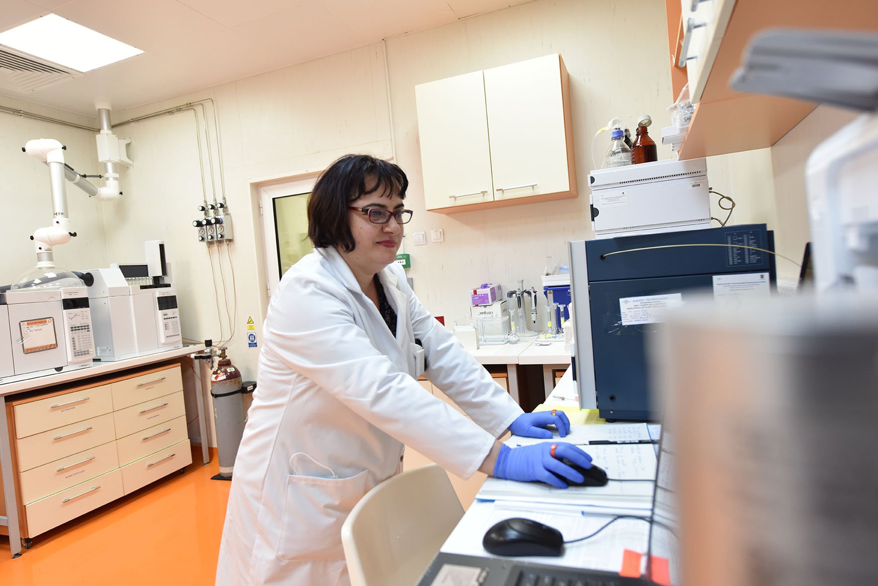
Every imaging agent is backed by rigorous analytics—radio-HPLC, IC, GC and TLC chromatography, ligand tracer assays and dynamic light scattering—so we know exactly what we are injecting and what it does.
Imaging & analytics
- PET & SPECT functional imaging, integrated with CT/MR as needed.
- Preclinical small-animal µPET/CT for in vivo biodistribution studies — at the TR-19 cyclotron facility.
- Radio-chromatography: HPLC, IC, GC, TLC with radio-detection.
- Ligand tracer assays for binding and specificity.
- Dynamic light scattering (DLS) for particle characterisation.
Why functional imaging
It reveals biology in action — catching disease before it reshapes anatomy.
3 · Treat it
Targeted radiotherapy — radiation delivered from within
The same idea that lets us see disease can be turned to treating it. Swap the imaging isotope for one that emits therapeutic radiation, and the molecule that once lit up a tumour now delivers a damaging dose directly to it—targeted systemic radiotherapy that spares surrounding healthy tissue.
This "see-and-treat" pairing is the heart of personalised nuclear medicine: image a patient to confirm the target is there, then treat with a matched therapeutic agent. Our full research pipeline carries each candidate from radiochemical synthesis and aseptic preparation through quality control and preclinical testing (in vitro and in vivo) toward clinical preparation and application.
Beyond the clinic
Where this capability reaches
Nuclear medicine
PET/SPECT imaging agents and targeted radiotherapies for earlier diagnosis and personalised treatment.
Pharmacology
Radiotracers to follow how drugs distribute, bind and clear in living systems.
Biology & agriculture
Nuclear techniques applied across biology, pharmacology and agricultural research.
Materials physics
Interdisciplinary use of radioisotopes and irradiation in materials research.