A platform, not a single technique
Matter under Radiation is the research direction built around TIESR—our hub for characterising materials and living systems across every scale, and for testing how they respond to radiation.
Most research questions are not answered by one instrument. They need a workflow: what does this sample look like? what is it made of? how does it behave when we irradiate it? TIESR brings all three under one roof, pairing a full ladder of imaging and compositional tools with the radiation sources of our accelerators — the TR-19 cyclotron and the 3 MV Tandetron.
That makes this direction the connective tissue of the department: the place where samples from our accelerators—and from external partners in materials science, biology, heritage and space research—are imaged, analysed and stress-tested.
See it & measure it
Imaging and composition from the millimetre to the atom
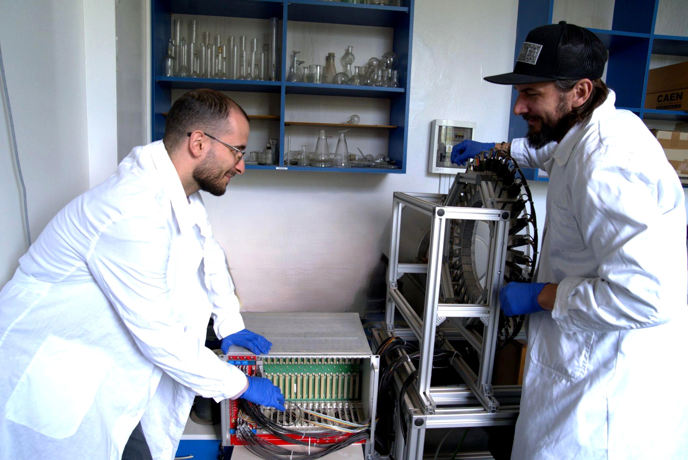
No single microscope spans every scale. TIESR's strength is the ladder: start with non-destructive 3D X-ray tomography to see internal structure, zoom into surfaces and microstructure with electron microscopy, then resolve individual features down to the nanometre with atomic force microscopy.
Alongside the imaging, a suite of elemental techniques answers what a sample is made of—from bulk down to trace concentrations—using X-ray fluorescence, laser-ablation ICP-MS and energy-dispersive spectroscopy.
Featured collaboration: imaging meets ion beams
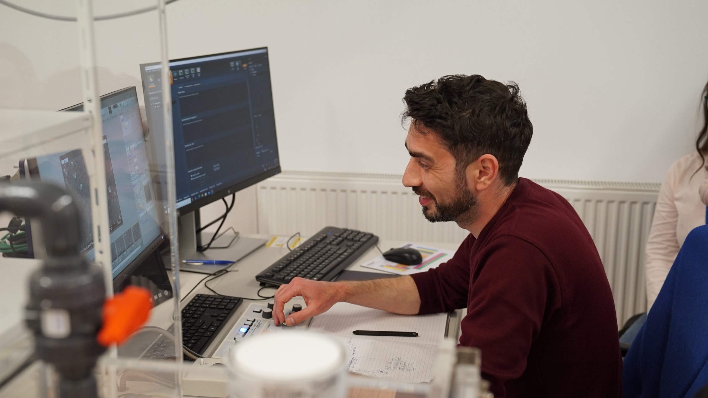
One DFNA study put two of the platform's techniques to work on the same samples — atomic force microscopy (AFM) to map surface topography and Rutherford backscattering spectrometry (RBS), on the institute's tandem accelerator, to measure composition and thickness. The team characterised indium-nitride (InN) and zinc-oxide (ZnO) thin films grown by magnetron sputtering: they measured mean surface roughnesses of about 12 nm (InN) and 27 nm (ZnO), resolved the films' InxN1-x and ZnxO1-x stoichiometry, and showed that the substrate temperature shifts the InN composition. A textbook case of the platform idea: see the structure, then measure what it is made of.
Published I. Burducea et al., Characterization of Indium Nitride and Zinc Oxide Thin Films by AFM and RBS, Rom. Journ. Phys. 58 (2013) 345–353.
A collaborative project Bringing together DFNA's ion-beam analysis (RBS) and atomic force microscopy at IFIN-HH, with the Faculty of Physics, University of Bucharest, and the National Institute for Optoelectronics (INOE 2000), which provided the nanomaterials.
Test it under radiation
The nuclear spine — how matter and life respond to radiation
Proton radiobiology
External proton beam line from the TR-19 cyclotron, with picoampere currents for controlled-dose irradiation of cells and biological samples.
Space-radiation simulation
Ion beams used to mimic the radiation environment of outer space, testing the resilience of materials and electronics for space applications.
Solid-target irradiation
Irradiation of solid targets for radioisotope production and radiochemical processing, supporting both research and medical applications.
µPET / CT imaging
Small-animal PET-CT for preclinical molecular imaging with sub-millimetre resolution—following radiotracers in living systems.
Complemented by NaI(Tl) and CsI(Tl) radiation spectrometry for β, γ and α detection.
One sample, one workflow
How the platform comes together
Image the structure
Non-destructive 3D X-ray CT reveals internal architecture; SEM and AFM zoom in on surfaces and microstructure down to the nanometre.
Map the composition
XRF, LA-ICP-MS and EDX identify what the sample is made of, from major elements down to trace concentrations.
Probe the radiation response
Controlled irradiation—with cyclotron and ion beams—tests how materials and living systems behave under radiation, then we re-image and re-analyse to measure the effect.
Close the loop
Before-and-after characterisation turns "what happened?" into quantitative, publishable results—linking structure, composition and radiation effect.
The connective tissue of DFNA
A shared platform that amplifies every other direction
For Archaeometry
Non-destructive imaging and elemental fingerprinting of artefacts and alloys—revealing how objects were made and where their materials came from.
For Ion Beam Applications
Characterising materials before and after ion-beam modification—closing the loop between making a material and proving what changed.
For Radiopharmaceuticals
Solid-target irradiation and µPET/CT preclinical imaging—bridging radioisotope production and the biology that proves it works.
For materials science
Multi-scale structure and composition for metals, ceramics, polymers, thin films and nanomaterials—including radiation-hardness testing.
For life sciences
AFM and electron imaging of cells, tissues and biomolecules, plus controlled-dose radiobiology on our accelerator beam lines.
For space & electronics
Simulating the outer-space radiation environment to test the resilience of components, materials and electronics destined for orbit.
CT — examples of our work
Volumetric reconstructions and slice views from the Nikon XT H 225 micro-CT at TIESR
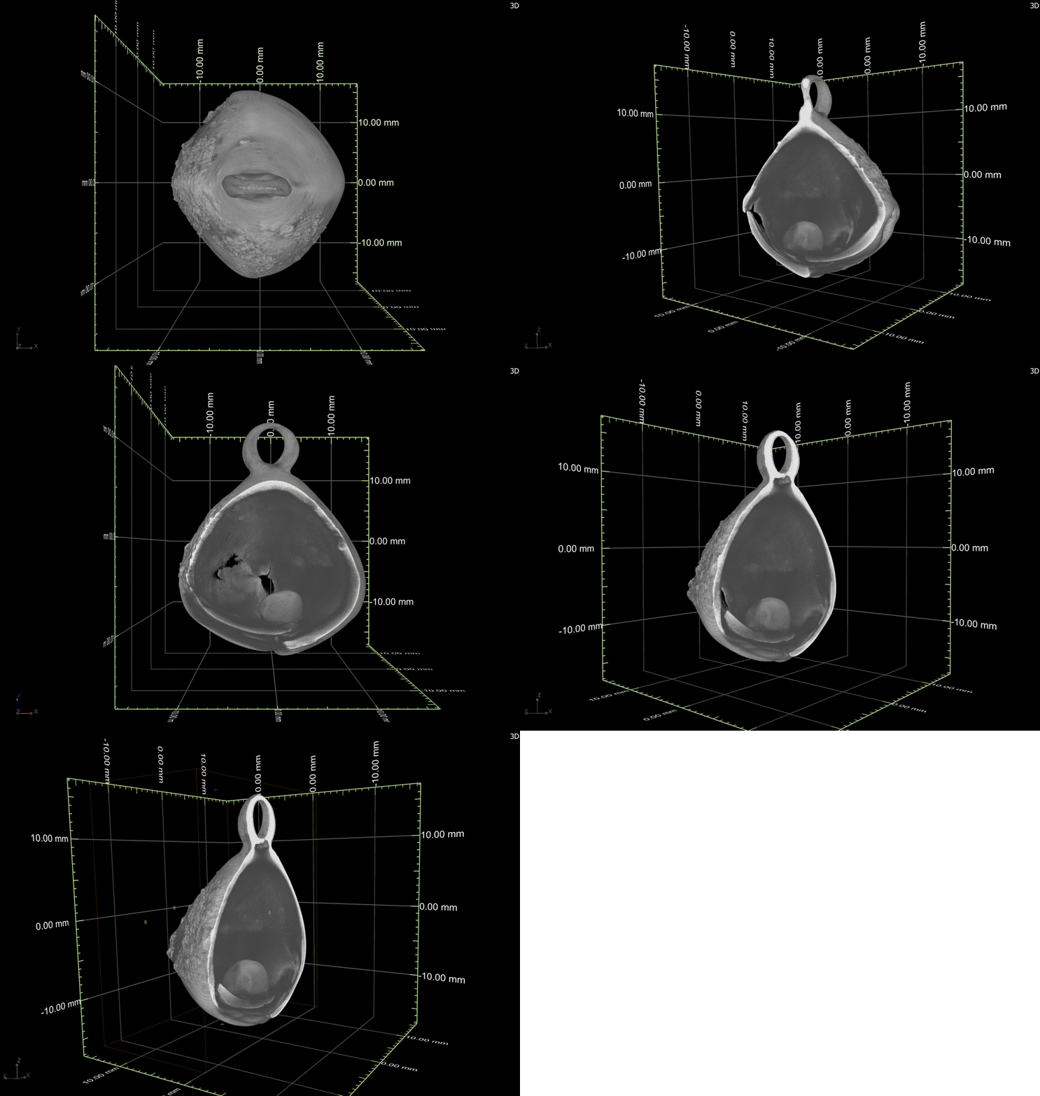
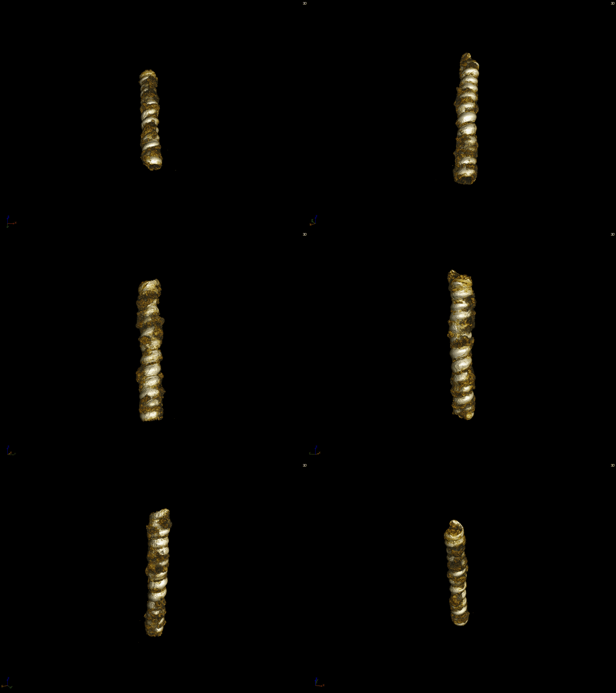
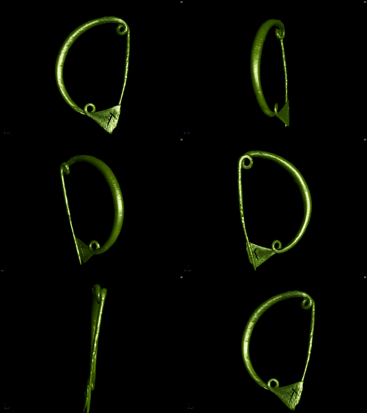
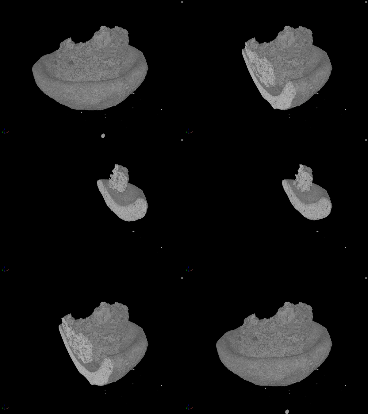
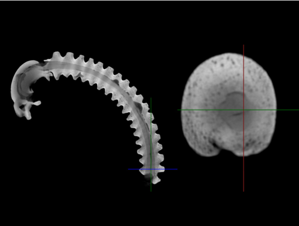
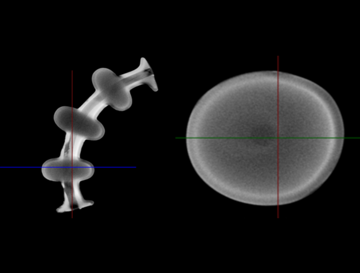
Reconstructed from micro-CT scans on the Nikon XT H 225 at TIESR. Click any image to enlarge.
AFM classic — examples of our work
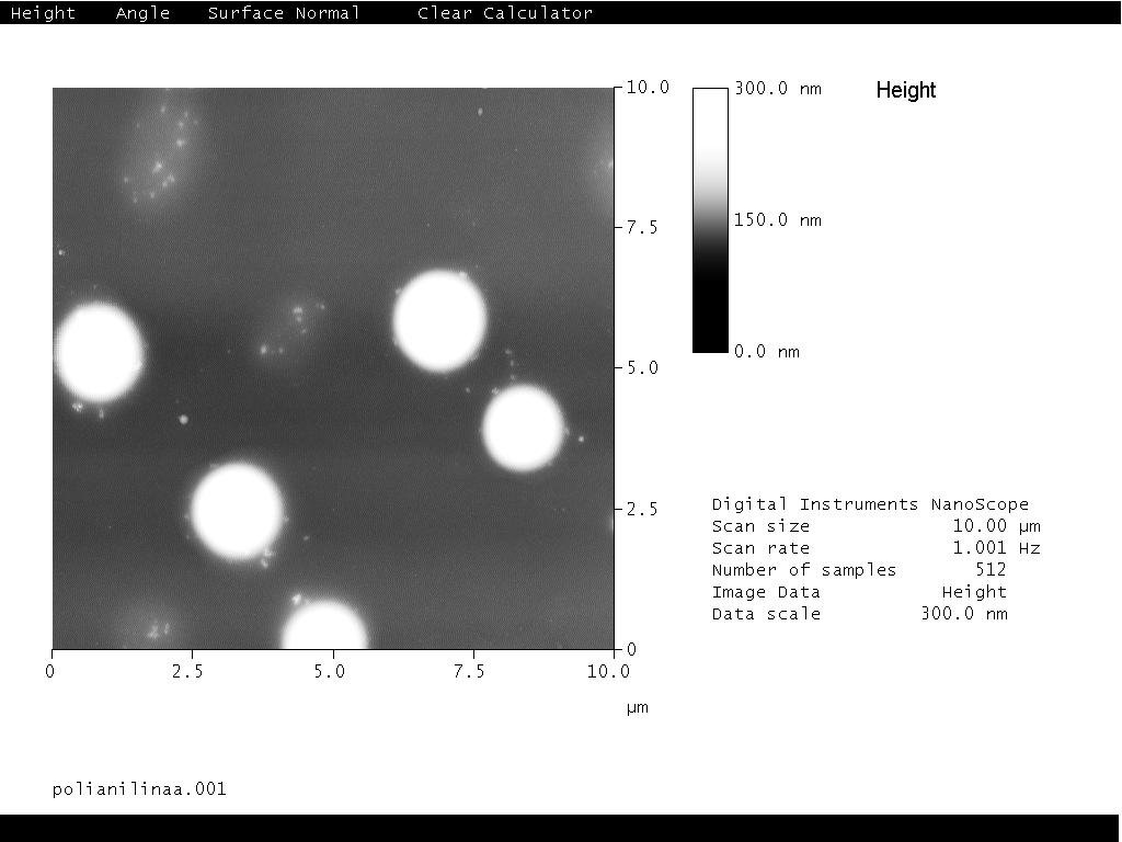
Representative AFM images acquired in the NANOMAT laboratory at TIESR
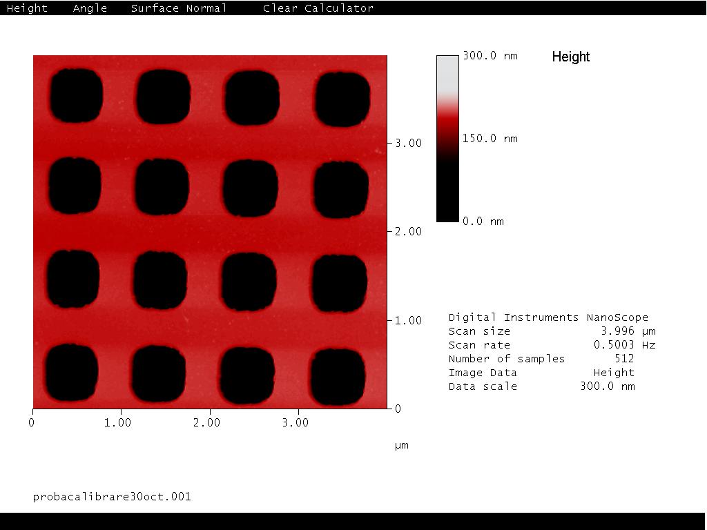
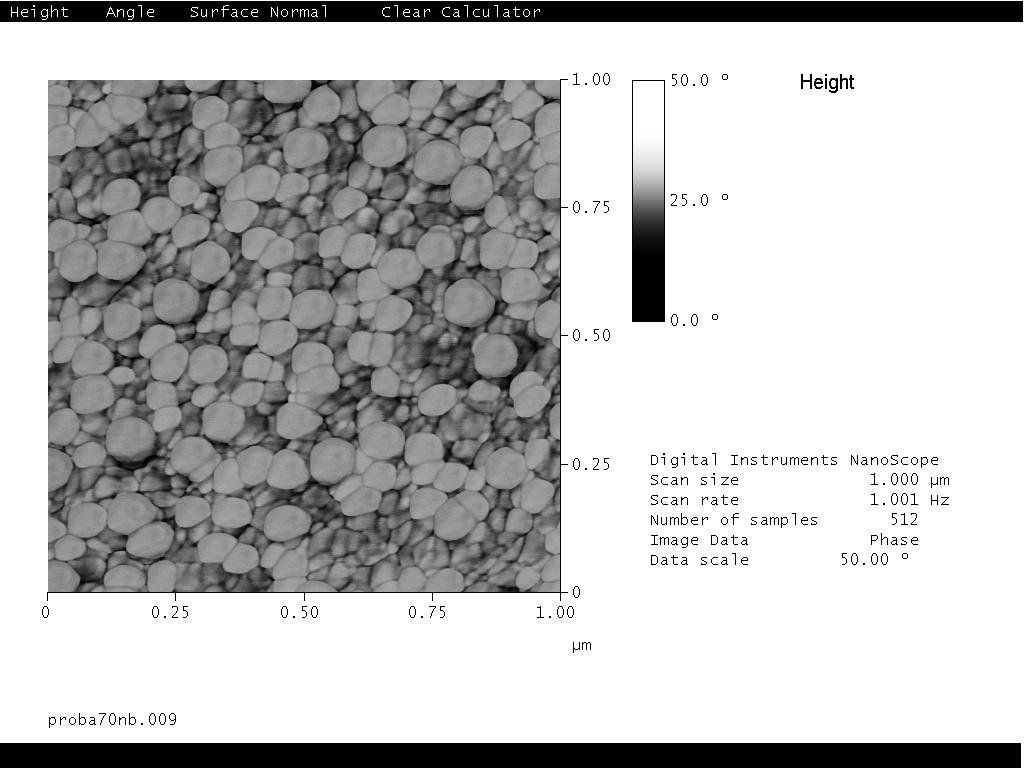
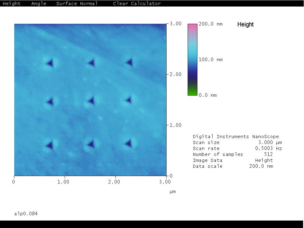
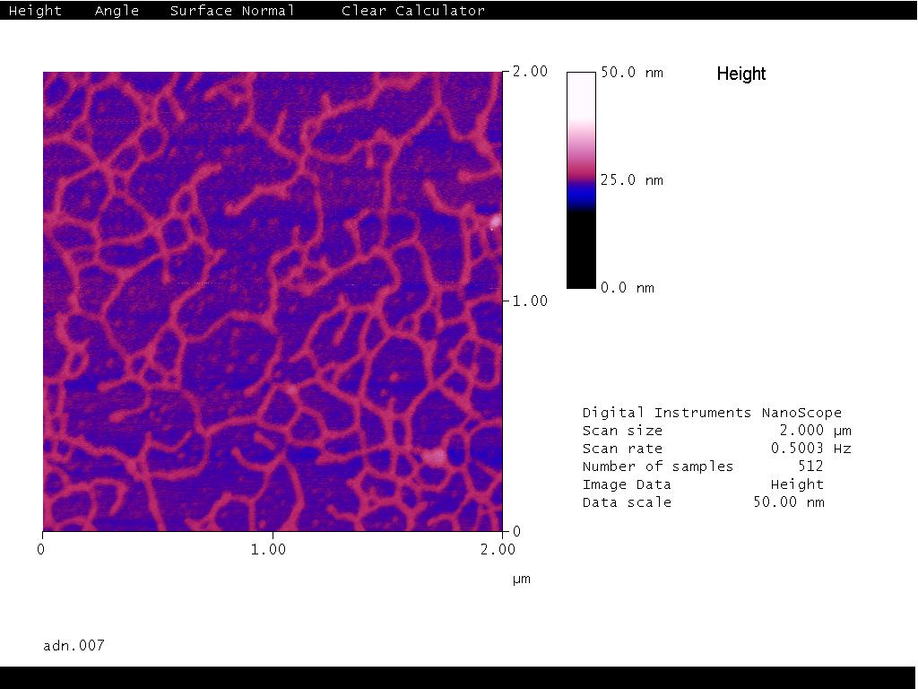
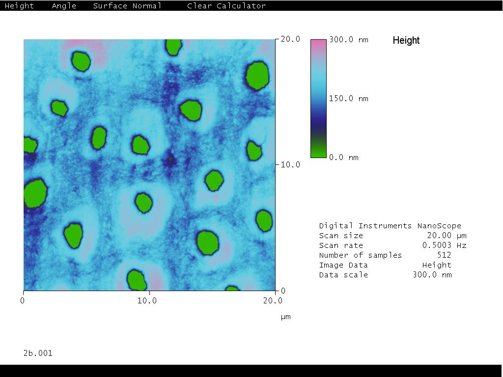
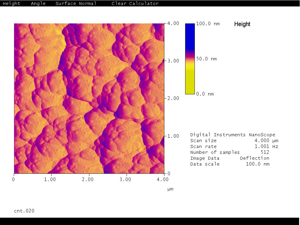
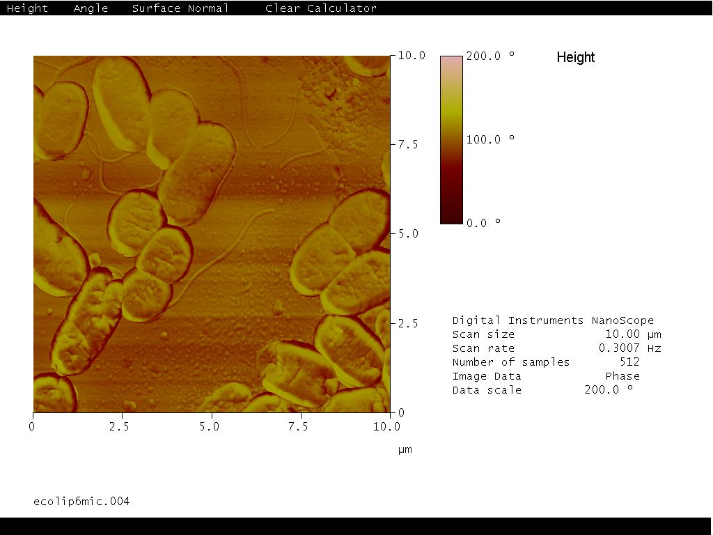
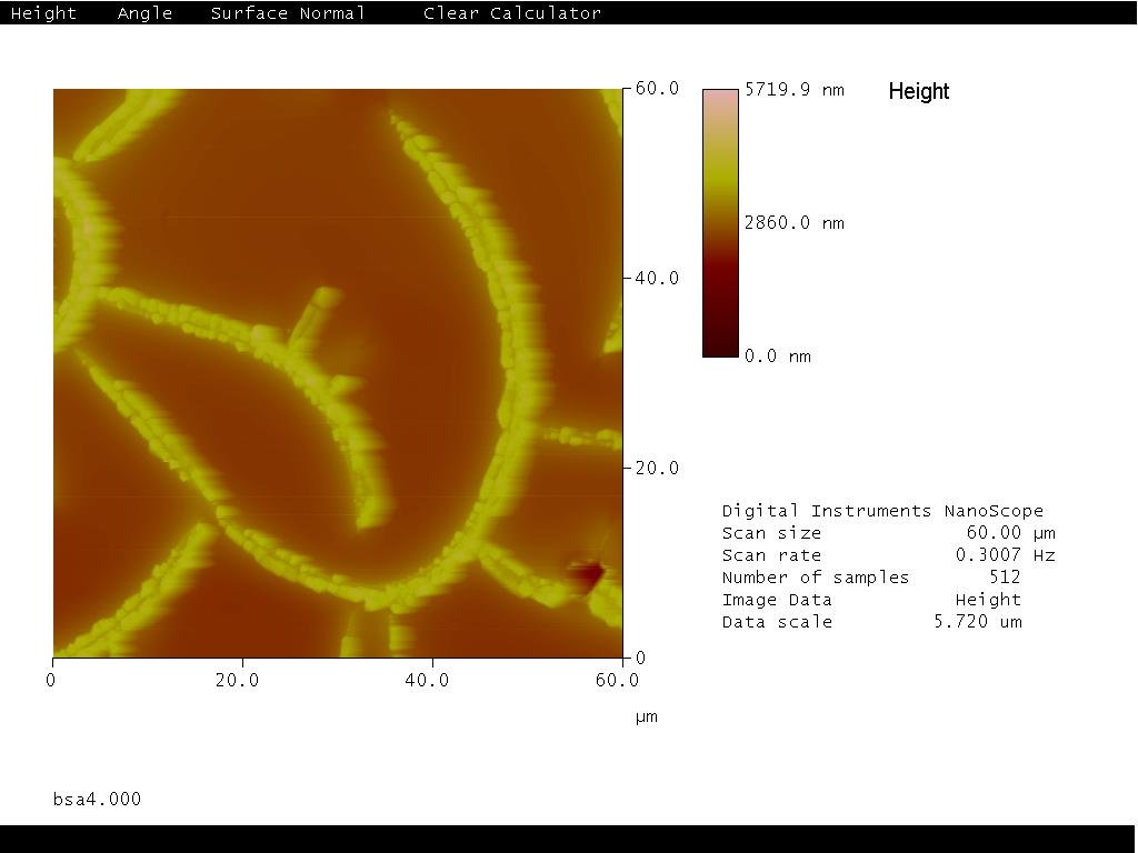
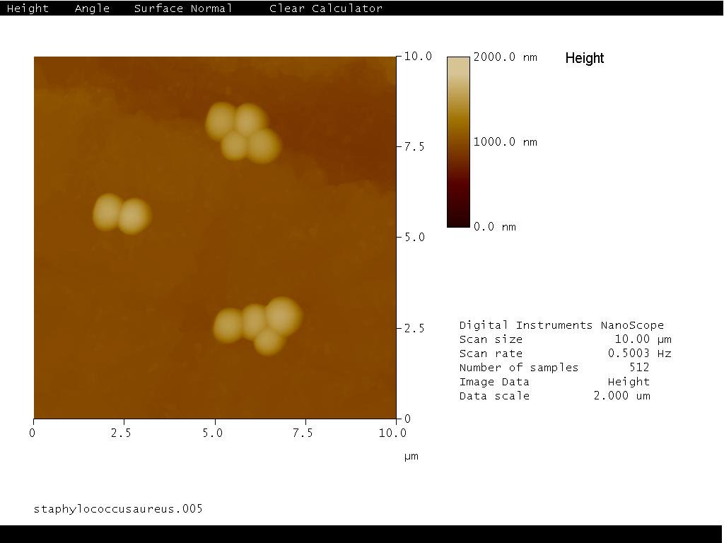
All images acquired with the MultiMode NanoScope III A Controller in the DFNA NANOMAT laboratory. Click any image to enlarge.
SEM — examples of our work
Surface imaging acquired on the Carl Zeiss Evo MA15 scanning electron microscope at TIESR
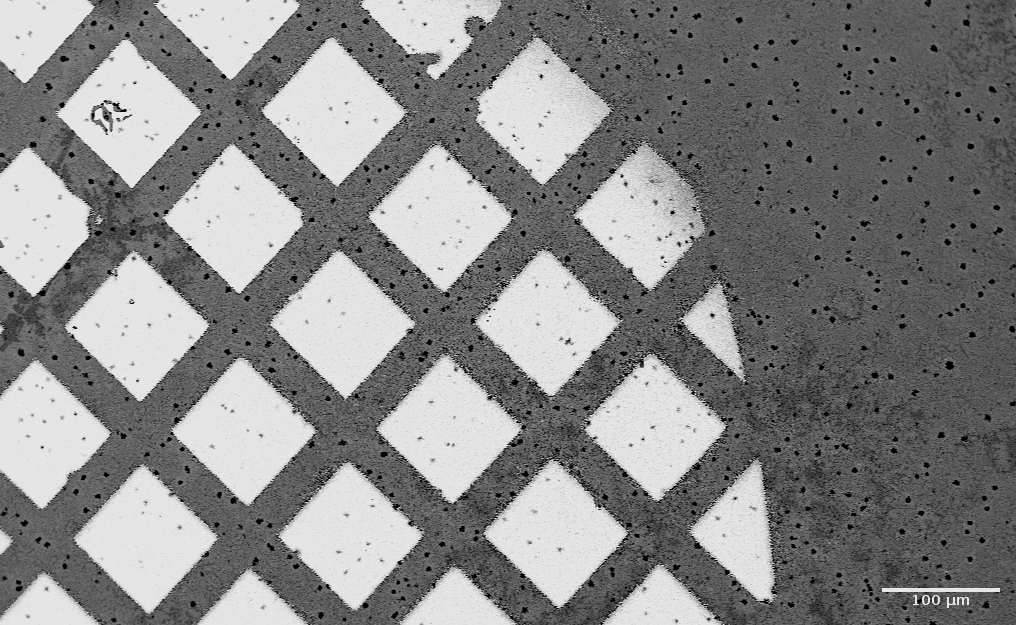
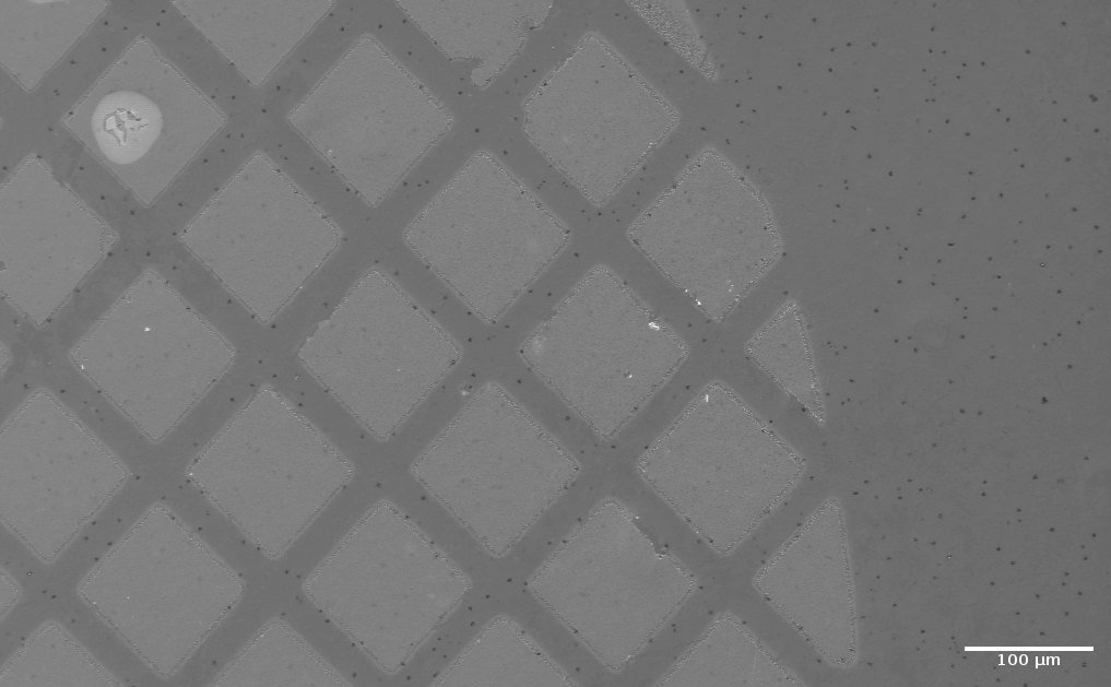
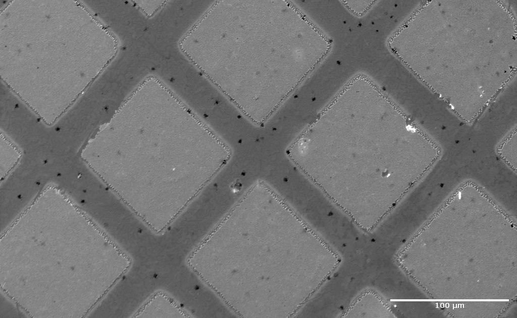
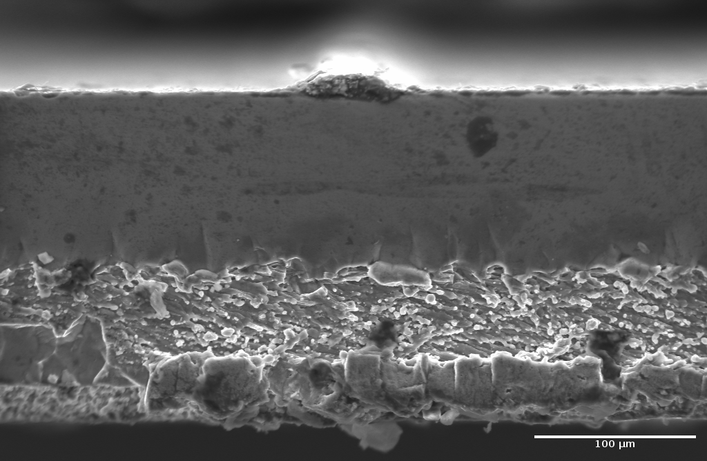
Acquired on the Carl Zeiss Evo MA15 SEM at TIESR. Click any image to enlarge.
Built on TIESR
Every capability in this direction lives in the TIESR facility—Testing, Trials and Experiments with Radiation Sources. Head there for the full instrument line-up, specifications and example results.
Whether you are a researcher who needs a sample imaged and analysed, a partner with a radiation-testing problem, or a student looking for a project, we would like to hear from you.
Explore TIESR Get in touch