Archaeometry can be defined as "the application of the exact sciences in the field of art and archeology." This interdisciplinary field is based on the use
of analytical techniques, initially developed in the field of materials sciences (techniques based on physical and chemical phenomena) in the study of art and archaeological objects. Studies can thus provide
historians and archaeologists with qualitative and quantitative information that can be used to understand ancient societies.
In DFNA we have a wide base of advanced experimental facilities that allow us to perform complex archaeometric analyzes (dating, determination of elementary
and imaging composition) using various analytical techniques (AMS, CT, XRF, PIXE, LA-ICP-MS, SEM) . The experimental facilities that can be used for archaeometric applications are the following:
• Tandetron type accelerators of 1 MV and 3 MV;
• Chemistry lab;
• X-ray computed tomography (CT) installation;
• Inductively coupled plasma spectrometer and laser ablation LA-ICP-MS (Laser Ablation - Inductive Coupled Plasma Mass Spectrometer);
• Stationary and portable X-ray fluorescence (XRF) spectrometers;
• Scanning electron microscope with EDX module (SEM);
• Optical microscopes.
For this type of research it is absolutely necessary to create an interdisciplinary community. Research in the field of archeometry involves the creation of networks
that create a close link between archaeologists, museographers and historians on the one hand and physicists and chemists on the other hand, who in turn come from various museums, cultural institutions and research institutes,
and which may be interconnected internationally. In recent years, a critical mass of researchers with relevant expertise in the field of archaeometry has focused on DFNA - in particular, C-14 dating (AMS), chemical composition
analysis (XRF, PIXE) and imaging (CT). This group is visible both nationally and internationally, being involved over time in numerous research projects.
More details about archaeometry can be explored on the study page cultural heritage în IFIN-HH.
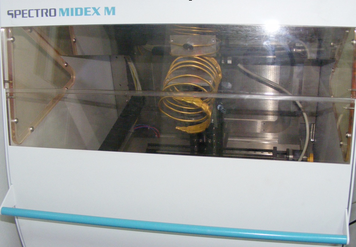 Spiral gold Dacian bracelet from a batch of 13 bracelets with composition: Au = 84.2-92.9%, Ag = 6.3-16.2% and Cu = 0.4-2.1%, analyzed in DFNA with the XRF Midex M spectrometer
 Fragment of anthropomorphic container, with polished front and covered with excised decoration and inlay with white paste, painted on the neck and face with red ocher.
Smooth and undecorated back. It comes from the Neolithic site from Vãdastra, south of Oltenia, about 5200-4900 Cal. BC. Collaboration with the Institute of Archeology "Vasile Parvan"
Fragment of anthropomorphic container, with polished front and covered with excised decoration and inlay with white paste, painted on the neck and face with red ocher.
Smooth and undecorated back. It comes from the Neolithic site from Vãdastra, south of Oltenia, about 5200-4900 Cal. BC. Collaboration with the Institute of Archeology "Vasile Parvan"
Responsible: Roxana Bugoi, Dragoș Mirea, Daniela Stan, Mihai Straticiuc
Analytical techniques based on atomic and nuclear phenomena can be used successfully in the study of objects of art and archeology, giving archaeologists
and museographers clues about the raw materials and manufacturing techniques that our ancestors had discovered and perfected. In some cases, markers / trace elements
and compositional patterns may reveal complex exchange / trade networks and the origins of the artifacts under study. Physico-chemical analyzes on heritage objects prove to be extremely
useful in the conservation and restoration processes, but also for testing the authenticity of artifacts proposed for purchase in museums or appeared on the art objects market - see the case
of the famous Dacian bracelets, expertized in and IFIN-HH (possibly links to articles). Cultural heritage assets are subject to degradation through 'aging', a process that must be understood,
both in its causes and in its dynamics, in order to find ways to prevent or slow it down.
• X-ray Fluorescence (XRF - X-ray Fluorescence) using fixed spectrometers (elementary laboratory analyzes) and portable (laboratory and in-situ analyzes directly in museums,
galleries, collections) allows the identification of chemical elements (from Mg to U ) present in the artefacts in a minimum concentration of tens of parts per million (ppm). The analyzed
surface has a diameter of 30, and the analyzed depth of the sample can vary between tens, up to hundreds of micrometers, depending on the analyzed material and the detected element. XRF is
par excellence a non-destructive technique, offering a multi-element quantitative analysis for a wide range of samples, from metal alloys to glass and ceramic artifacts.
• The ion beams accelerated using Proton Induced X-ray Emission (PIXE) at the 3 MV Tandetron particle accelerator, allow the identification and quantification of chemical elements from
Na to U, present in concentrations of the order of ppm in the analyzed objects. With the help of a 3-axis sample positioning system, which moves with micrometric precision and using a proton
beam with a diameter of ~ 1 mm, the artifacts can be analyzed both in vacuum and in He atmosphere, at atmospheric pressure. The probe depth is of the order of tens of microns and, as in the
case of the XRF method, it depends on the density of the analyzed material and on the chemical element whose concentration is to be determined.
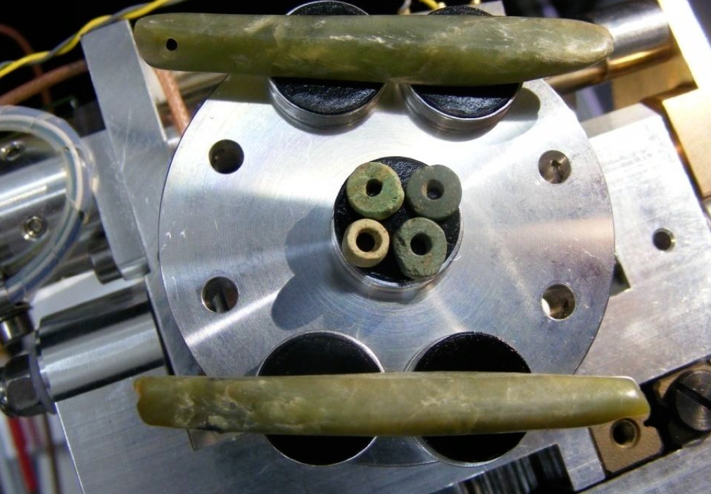 Neolithic ornaments (Gumelniţa civilization) made of nephrite positioned on the test stand of the SEM-EDS ZEISS EVO MA15 installation
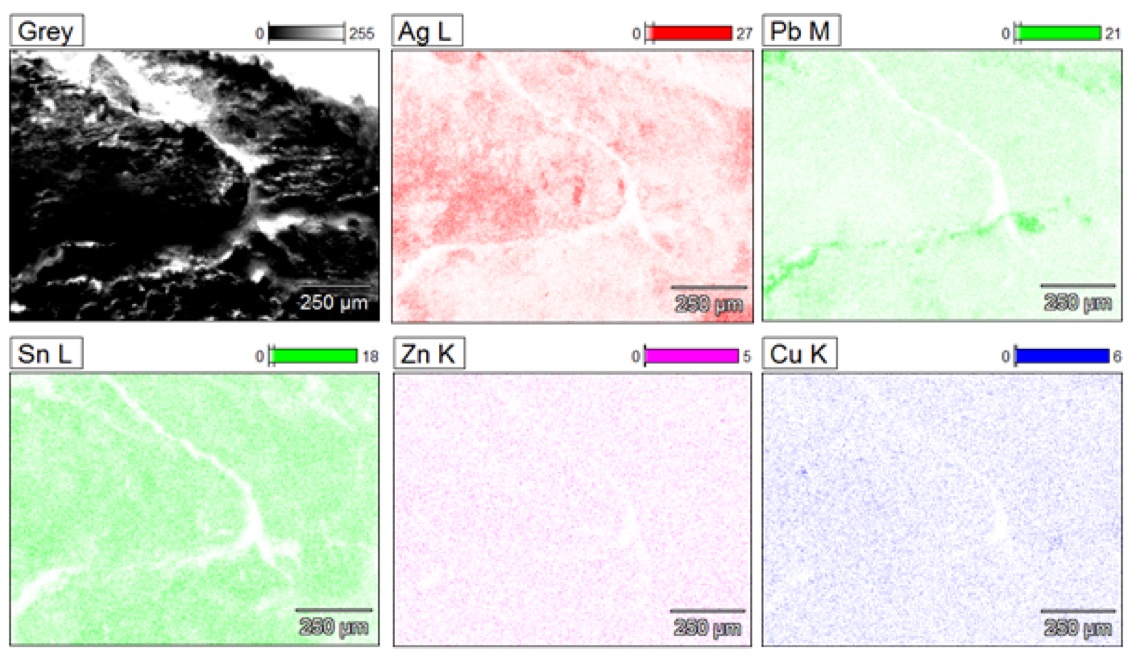 Elemental map made with SEM EDS on Dacian and Roman silver coins, dating from the 1st century BC. - III d.H. (Histria, Dobrogea)
Responsible: Paul Mereuță
Within DFNA, the SEM-EDS analytical technique is applied for the study of a diverse range of materials. Due to the possibility of optimizing the parameters of the electron beam
(E = 1-30 keV, I = 1-2000 nA) this method is non-destructive, but involves some geometric limitations imposed by the fact that the analyzes can be performed only in vacuum. The resolution of the SEM image can reach up to 3 nm, while
the energy resolution of the X-ray detector (EDS system) is 129 eV (K? Mn), with a detection limit of about 100 ppm. Thus, the chemical elements from C to U can be detected and those with an atomic number greater than 10 can be quantified.
At the same time, two-dimensional elemental maps can be obtained for certain portions of the analyzed artifacts.
References
[1] P. Mereuta et al., SEM-EDS AS INVESTIGATION TOOL FOR ARCHAEOLOGICAL ARTIFACTS – THE CASE OF NEPHRITE ADORNMENTS, Romanian Reports in Physics 71, 802 (2019)
[2] D. Cristea-Stan et al., ANCIENT SILVER AND BRONZE METALLURGY STUDIES
BY MICRO-PIXE AND SEM-EDS, Romanian Journal of Physics 63, 204 (2018)
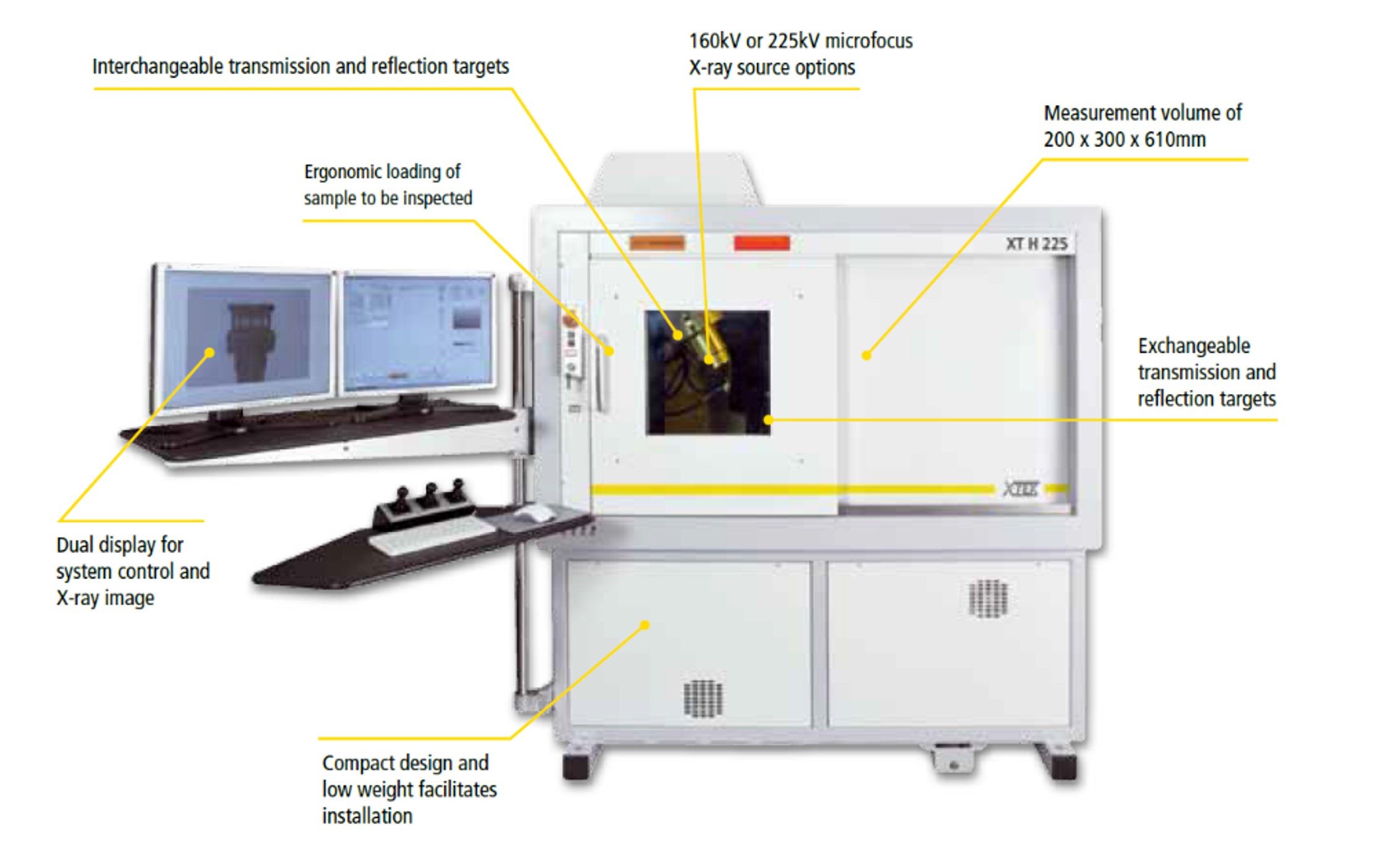
X-ray Computerized Tomography system Nikon XT H 225
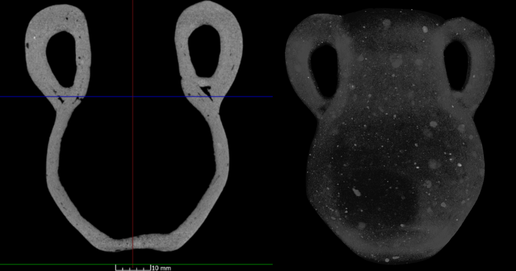 Ceramic amphora with damaged body (Costisa), frontal section (left), highlighted clay inclusions (right)
Anywhere the internal structure matters, X-ray and CT technology serves as an efficient tool to provide valuable information. Detailed capture and measurement of internal features is often vital for quality control or defect analysis. Besides these features, the versatile computerized tomography system accomplishes maybe the most important task: non-destructive material analysis. This enables a wide variety of material research across various industries. Archaeometry is one of the areas of interest where archaeologists need information as: object manufacturing technique, casting quality of metal parts, material's degree of porosity, density, size and orientation of the inclusions in the material, specific characteristics that enables connections between different archaeological sites, etc. Such observations are possible through computed tomography thus highlighting the potential of this technique in the study of archaeological artefacts.
Our department's X-ray computed tomography system is supplied by Nikon Metrology, model XT H 225. Equipped with a powerful micro-focus X-ray source (3 μm focal spot) and a detector with a high spatial resolution of 1900 x 1516 , with a pixel size of 127 μm, this equipment ensures a high speed image acquisition at the highest quality. Depending on the type and density of the investigated material, but also on its thickness, the working parameters of the tomograph can be adjusted so that we can capture as many inner details as possible. The system operates between 30 and 225 kV, at a maximum current of 1 mA, with the possibility of using aluminum or copper filters that attenuate low energy radiation. The tomograph also contains a precise object positioning system that allows movements of the sample in all three directions, as well as 360o object rotations around its own axis, but also inclination movements by ±30o. With the help of a high-performance software, VGStudio MAX 3.0, the data collected by the tomograph are subsequently analyzed and processed, practically building a complete three-dimensional representation of the object from numerous two-dimensional projections acquired by the detector.
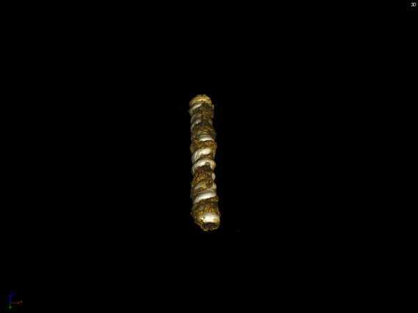
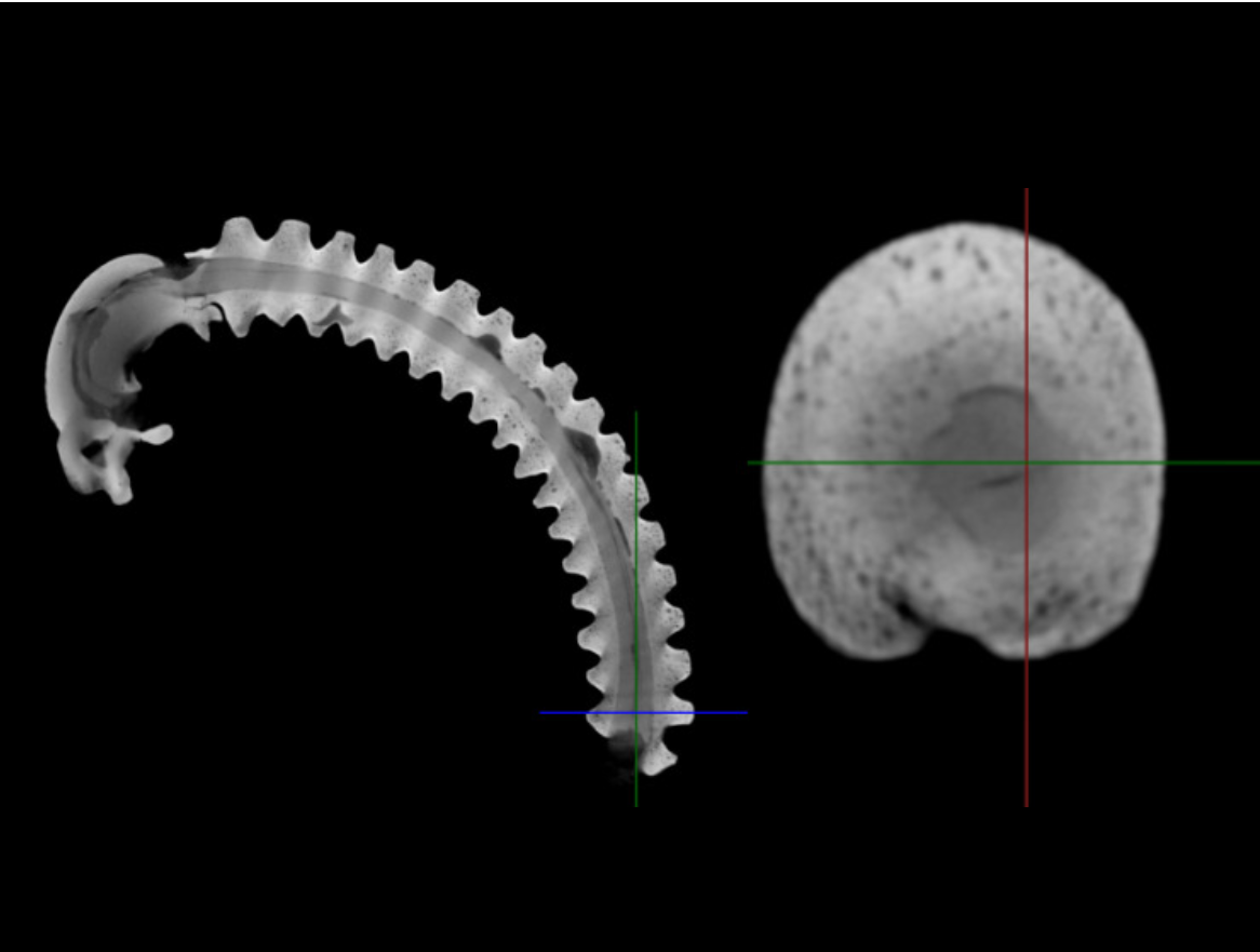
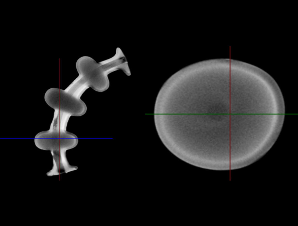
Fibula from Oltenia (inv. No. 08154) with a ribbed aspect of the spring: longitudinal section (left), cross section (right)
Fibula from Oltenia (inv. No. 08153) with nodules on the spring: longitudinal section (left), cross section through one of the nodes (right)
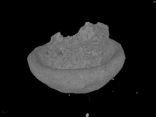
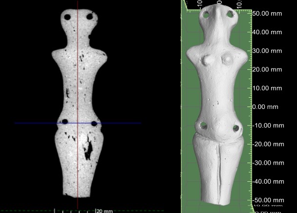
Anthropomorphic statuette, front section (left), three-dimensional representation of the object (right)
Clopotel-pandantiv metalic (Targusoru Nou)
Vas mic ceramic cu pamant
References:
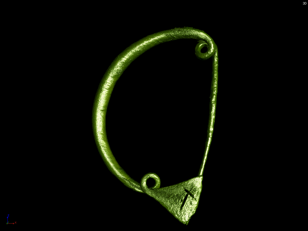
[1] Cărăbişi V.-Şt., Popescu A.-D., Petruneac M.,Focşăneanu M., Cristea-Stan D., Constantin F., 2020. Early Iron Age fibulae from Balta Verde, Romania: typology, combination and manufacture. In: Mărgărit, M, Boroneanţ, A. (eds.), Beauty and the eye of the beholder. Personal adornments across the millennia. Cetatea de Scaun, Târgovişte, pp. 413-429
[2] Popescu A.-D., Cărăbişi V.- Şt., SÎrbu R., Petruneac M., Cristea-Stan D., Focşăneanu M., Mihălceanu S., Constantin F., Tehnici de realizare a două fibule din prima epocă a fierului descoperite În Oltenia, Studii şi Cercetări de Istorie Veche şi Arheologie 71.
[3] Alin Frînculeasa et al, Turanici la Dunărea inferioară - complexe cercetate recent în nordul Munteniei, Materiale şi cercetări arheologice (serie nouă), XVI, 2020, p. 199-228
.gif)